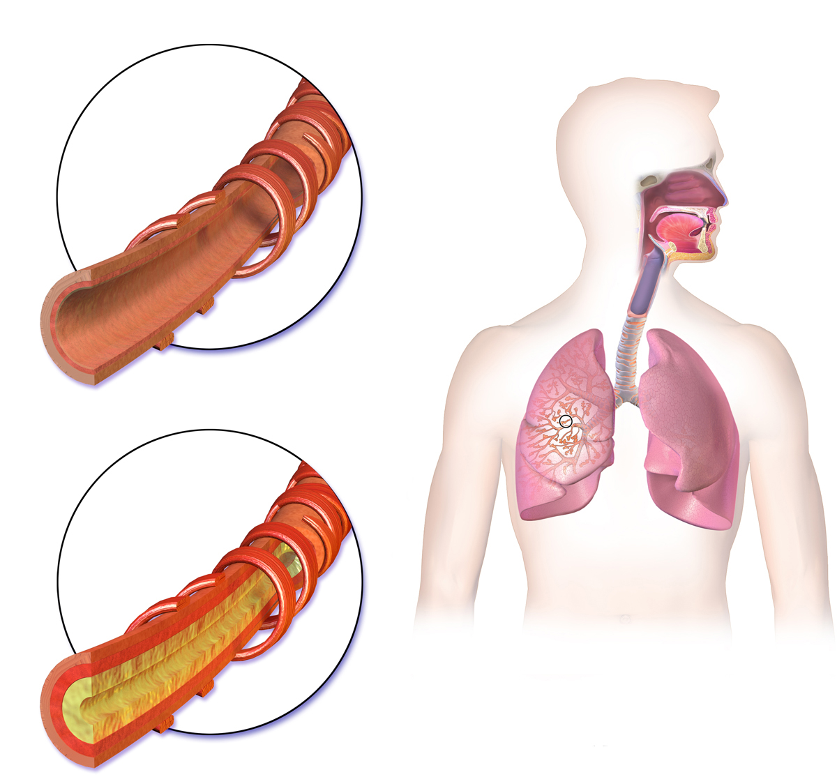
Symptoms
In most cases, the source of the bleeding is in the bronchi, and the percentage of these cases is 90%. Hemoptysis cases in which the source is in the pulmonary system account for a total of 5% of all cases. The pulmonary system is responsible for carrying de-oxygenated blood away from the heart to the lungs and returning blood, which is oxygenated from the lungs back to the heart. There are many risk factors associated with Hemoptysis, like bronchitis, trauma, and tuberculosis.
Diagnosis
For the diagnosis of the symptoms of Hemoptysis, the first step would be a chest radiograph. A total of 30% of the suspected cases appear to be confirmed after the radiograph results. Another useful approach to diagnose Hemoptysis is bronchoscopy, in which a medical visualization device is inserted in the airway. The doctor can also use CT to identify the bleeding source and to find the cause of it.
Treatment
For massive hemoptysis treatment, there is bronchial artery embolization. In this process, there is an introduction of an object in the body of the patient, which is done by an interventional radiologist. An interventional radiologist takes help from fluoroscopy guidance. He inserts a catheter in the person’s femoral vein, located in the upper thigh. After this, the radiologist guides the catheter into the bronchial artery and injects embolization materials into the branch of the bronchial artery, which is causing the bleeding. The embolization material blocks the bleeding after the successful completion of the procedure. After the process of embolization, the person has to undergo angiography to determine whether the operation was a success or not.
There are high rates of success in this invasive clinical treatment process. Still, if the bleeding is recurring, the embolization process can be done again. Due to Hemoptysis, the patient may experience many difficulties like chest pain and difficulty swallowing. The most serious problem is inflammation of the spinal cord, but the chances of this happening are only in 1.4%-6.5% of the cases.
Surgical treatment options are also available. But these options are for some exceptional cases only, where the embolization process is not successful, or if the condition appears again after many embolization processes. The mortality rate of surgery is 14%-18%.
Doctors can also go with other treatment procedures like Endobronchial Tamponade. In this process, an absorbent plug is used to stop the bleeding, and it is the best option for those patients who are not appropriate for surgery or embolization processes. Another minimally invasive approach is rigid bronchoscopy. This process can be used to treat the patients who have breathing problems, or their blood flow is impaired. Bronchoscopy can also help to clear the airway and help the physicians to do further treatments.
Dr Nikolas Charalambous is named among the most prominent interventional radiologist. Get a free consultation for a minimally invasive treatment of brain, neck, and spine.

Andrea Avraamidi 55-57, Strovolos 2024, Nicosia,
Cyprus
Email: consulting at ncir.com.cy
Phone: +357 99 44 08 22