Symptoms
The location and size of the intracranial arteriovenous malformation (AVM) will define the symptoms you are observing. There is also a chance that you may not feel any sign. In some cases, it is also possible that you may experience a headache that won’t go away. Other symptoms include weakness or numbness and seizures. There is a chance that you may feel continuous buzzing sound in your head. If the arteriovenous malformation (AVM) starts to bleed, there would be a chance of developing symptoms of less common nature, such as acute stroke or subarachnoid hemorrhage.
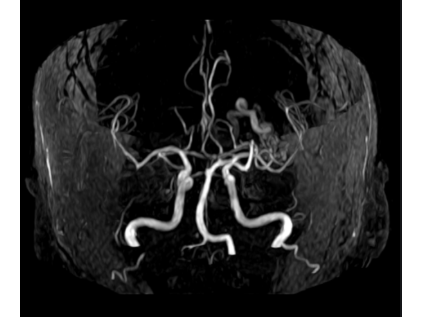
Diagnosis
The doctor can diagnose the condition by the help of medical imaging techniques, such as by taking an MRI or CT scan of your brain. To make the optimal and best treatment for the patient, the doctor may need to do a cerebral angiogram (or DSA) using the X-Ray guidance which involves the use of a contrast material directly injected in the patient’s brain.
Treatment
For the treatment of the arteriovenous malformation (AVM), there are three possible options:
The first is endovascular embolization, the second is surgical removal, and the third is stereotactic radiosurgery.
The size, the location and whether there is any bleeding or not of the arteriovenous malformation (AVM) will define the best and optimal treatment plan. Commonly, the patient who is facing this condition may undergo more than one surgery or treatment plan.
If you go through an endovascular embolization, here is what you will experience. In this process, the radiologist will guide a microcatheter to the location of arteriovenous malformation (AVM) through an artery in your groin. After reaching the targeted area, he will plug the abnormal blood vessels with the help of medical-grade glue.
There is a form of radiotherapy, the stereotactic radiosurgery which is also used for the treatment of AVM. In this process, the arteriovenous malformation (AVM) is exposed so that the beams of gamma rays can be focused on it. As a result, the rays will damage the abnormal blood vessels, and it will make the flow of blood slower. Thus, the AVM will shrink, and over the course of months and years, it will turn into a scar.
For these procedures, you need to stay in the hospital for as long as required and recommended by the doctor.
Dr Nikolas Charalambous is named among the most prominent interventional radiologist. Get a free consultation for a minimally invasive treatment of brain, neck, and spine.

Andrea Avraamidi 55-57, Strovolos 2024, Nicosia,
Cyprus
Email: consulting at ncir.com.cy
Phone: +357 99 44 08 22