Symptoms
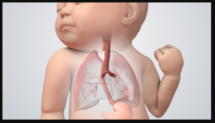
If the TEF is present from birth, its symptoms can be found during the prenatal ultrasound. The ultrasound can indicate more than average amounts of amniotic fluid in the amniotic sac, no fluid in the stomach, smaller abdomen and enlarged gullet area. The infants with TEF emit a huge amount of oral secretions; their skin can be bluish, and they also have possible respiratory distress, cough, gag and vomit.
The patient who has developed this condition and has been ventilated may experience the unexplained weight loss, repeated failure to wean and recurrent chest infections. If the person is non-ventilated, the symptoms may include pneumonia, repeated respiratory tract infection, hoarseness, fever, coughing up blood, difficulty in swallowing, shortness of breath and chest pain. The pain can be similar to a person who has cancer, along with some additional symptoms caused by the tumour.
Diagnosis
TED can be diagnosed through the usage of imaging techniques. If the doctor thinks your unborn baby can have TEF, a foetal MRI is the best way to diagnose it. Chest radiology is usually used for diagnosing the TEF in the infant born with TEF.
If the person is suffering from TEF and a nasogastric tube is inserted, it will be impossible to pass the tube into the stomach; instead, the tube will remain in the middle section of the chest cavity. CT radiography can also be used to diagnose the TEFs, but this method is not commonly used. Sometimes the contrast-enhanced studies are used to confirm the diagnosis. This technique can also be used to find the location, identify its size and direction. TEFs, which is acquired, can also be diagnosed through direct visualization with the usage of a metal tube with an attached camera.
Treatment
Surgery is used to treat the TEF. If an infant is in good health and has no complication along with the TEF, a primary repair can be done in the first few months after birth. If the patient’s birth weight is low, the repair can be delayed to avoid complications like pneumonia and other condition. The treatments of these patients should be conservative with the use of intravenous feeding, in which a tube is inserted through the skin directly in the upper pouch suction and stomach. This process is done until the risk is low so that the procedure can be done.
There should be some precaution be taken so that there would be no leakage or damage in the gullet. These precautions can be the placement of tracheostomy or endotracheal. To reduce the reflux, a gastrostomy tube can also be applied.
If the cancer is the cause of the TEFs, those can be inoperable. In these kinds of cases, a stent can be placed in the gullet so that the TEF can be isolated to prevent food and other material from entering the windpipe or lungs.
Dr Nikolas Charalambous is named among the most prominent interventional radiologist. Get a free consultation for a minimally invasive treatment of brain, neck, and spine.
Andrea Avraamidi 55-57, Strovolos 2024, Nicosia,
Cyprus
Email: consulting at ncir.com.cy
Phone: +357 99 44 08 22