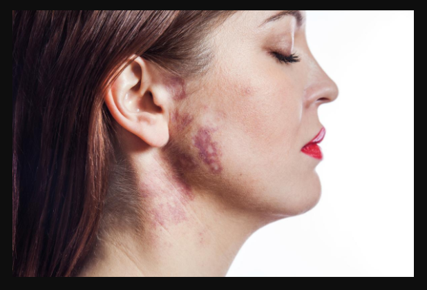
Symptoms
The primary and most common symptom is pain. Patients may feel symptoms depending on the location of the vascular malformation. The symptoms may be in the form of birthmarks and swelling of the limbs. In a patient who is suffering from lymphatic malformation, there can also be complications due to the infection. Some cases of arteriovenous malformation can be stressful on the heart, and they can even cause bleeding complications. If the person is suffering from arteriovenous malformation, he may experience a supply of low oxygen, shortness of breath, coughing, and fatigue.
Diagnosis
Most of the time, vascular malformation can be seen in physical examination. Ultrasound can be used to evaluate the superficial vascular lesions, but for the deeper lesions, ultrasound provides you with limited information. Ultrasound with sonography can be used to visualize the speed and the flow of the blood.
For evaluating vascular malfunction, MRI is the best diagnostic tool. MRI is so useful that it can show vascular malfunction’s core’s exact location; the core is called nidus. It can also confirm from where the malformation extends and what is the connection of it with surrounding vessels. MRI can also be used to gain more information on blood flow in the lesions, and also can be used to determine the success of the treatment during the checkup.
Minimally invasive methods like angiography and phlebography are used under medical imaging fluoroscopy. To help with the visualization of vascular malfunction, contrast material is injected. These methods are usually used before treatments like embolization and sclerotherapy.
Treatment
If the patient has mild symptoms, then conservative treatment is a good option. It is not always an option to perform surgical removal of vascular malfunction as this condition has a high rate of recurrence.
The treatment of choice for the vascular malfunction are minimally invasive techniques that are guided by imaging done by the radiologist. The methods which are used are sclerotherapy and embolization. These methods are done under fluoroscopy guidance. In these processes, the radiologist will guide a catheter into the vascular malformation, and small beads, alcohol, or glue will be inserted in the surrounding vessels so that the lymph and blood will be blocked to the malfunction location. The goal is to destroy the core of the malfunction. Angiography is performed to confirm the success of the process. Patients may be under local anesthesia for this process and may remain in the hospital for one day. There can be minimal discomfort for some days.
Dr Nikolas Charalambous is named among the most prominent interventional radiologist. Get a free consultation for a minimally invasive treatment of brain, neck, and spine.
Andrea Avraamidi 55-57, Strovolos 2024, Nicosia,
Cyprus
Email: consulting at ncir.com.cy
Phone: +357 99 44 08 22