Symptoms
If the person is suffering from this condition, he may feel the primary symptom - a moderate to severe back pain, which will worsen due to movement.
If the spinal cord is affected by the fracture, the person may feel tingling, shock in the affected area, numbness, weakness, and bladder or bowel dysfunction.
If the injury is in the middle or lower spinal cord, it is likely that it will affect the functionality of the nerves, the spinal cord, and even the brain, along with lower extremities and genital areas.
Diagnosis
The diagnosis of this condition will be made on the basis of full physical examination, which includes laboratory tests, testing reflexes, and medical imaging.
If the fracture is due to major trauma, there will repeat blood tests to see the circulation of blood.
If there is a metastatic bone disease, involved in vertebral fracture, there will also be a calcium test to see the level of calcium in the blood.
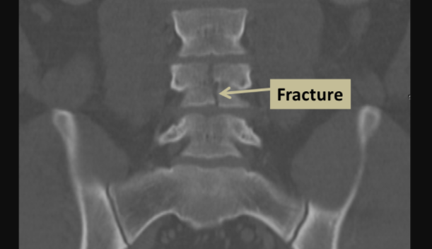
Imaging processes can also be used for diagnosing purposes. For the screening of spinal fracture, plain radiographs can also be used. It is very difficult to identify hairline fractures and non-displaced fractures. Such fractures in which the fractured bone pieces are still aligned cannot be diagnosed by using the spinal X-rays alone. Thus, the CT will be a good option to identify the condition and its extent.
MRI is the best option to identify the damage which has been done to the spinal cord and if the bone has become swollen. MRI is the best tool to identify the lesions in the bone and the spinal cord.
Treatment
If the condition is minor, the treatment will be non-surgical such as, spinal orthotic vest. It is a vest that is used to stabilize the spine.
If the condition is unstable, severe, or the effect has gone to the spinal cord, there will be a surgical treatment and stabilization of the spine so that there would be no further deformity. You may also have to take steroids depending upon what is the nature of the fracture.
There also some minimally invasive methods like Kyphoplasty and Vertebroplasty. In this process, cement is inserted into the area which is surrounding the fracture. The purpose is to stabilize the condition and reduce the pain.
Dr Nikolas Charalambous is named among the most prominent interventional radiologist. Get a free consultation for a minimally invasive treatment of brain, neck, and spine.

Andrea Avraamidi 55-57, Strovolos 2024, Nicosia,
Cyprus
Email: consulting at ncir.com.cy
Phone: +357 99 44 08 22